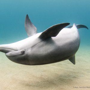
A new article from our team:Toxoplasma gondii has been described in several marine mammals around the world including numerous species of cetaceans, yet infection and transmission mechanisms in the marine environment are not clearly defined.
Abstact:
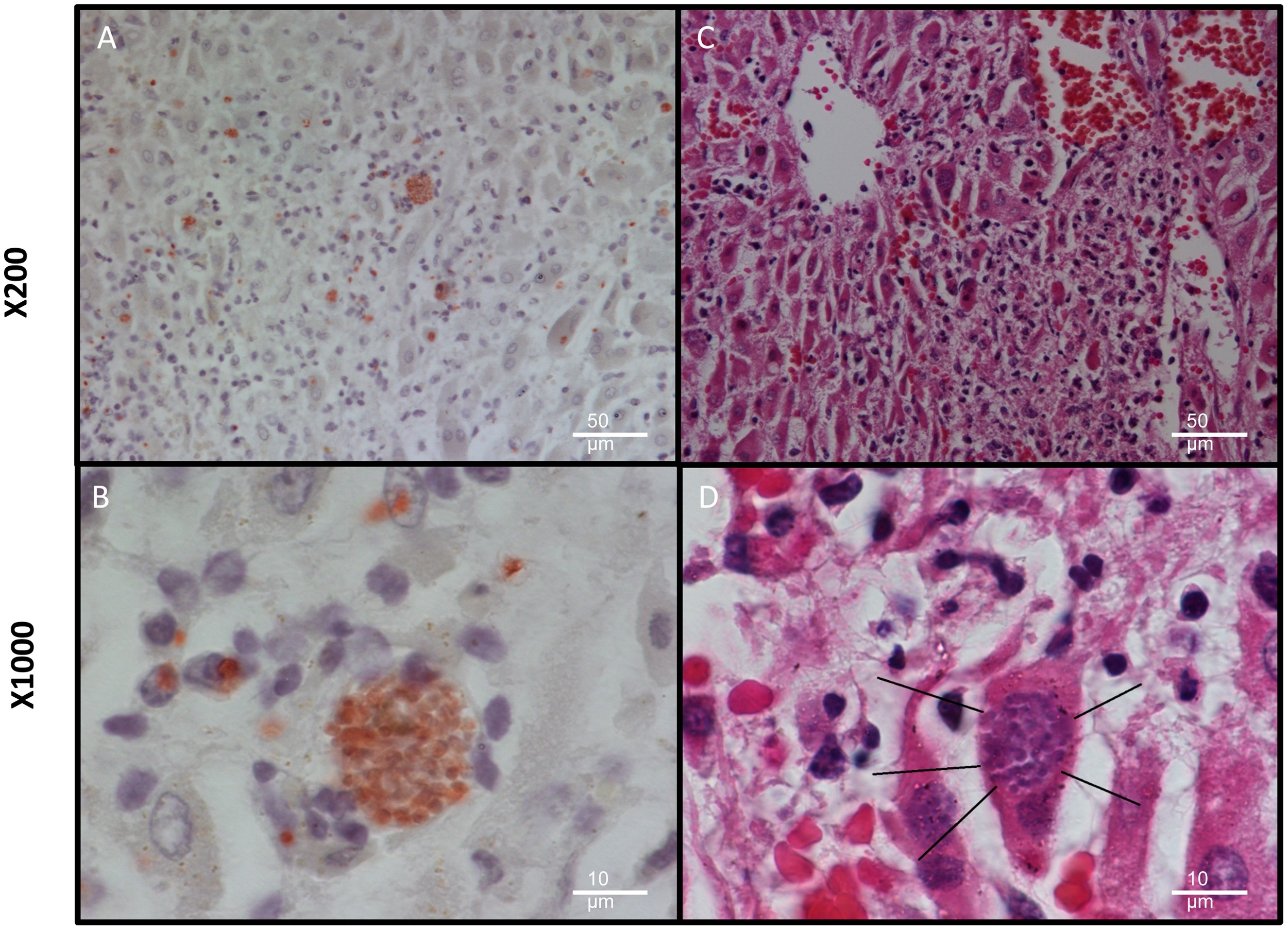
Toxoplasma gondii has been described in several marine mammals around the world including numerous species of cetaceans, yet infection and transmission mechanisms in the marine environment are not clearly defined. The Israel Marine Mammal Research and Assistance Center has been collating a database of all marine mammal stranding events along the country’s national coastlines since 1993. In this study, we describe the molecular detection and characterisation of T. gondii in three common bottlenose dolphins (Tursiops truncatus) including one case of coinfection with herpesvirus. The animals were found stranded on the Mediterranean coast of Israel in May and November 2013. In one of the three cases, the dolphin was found alive and admitted to intensive care. To our knowledge, this is the first report of T. gondii infection of marine mammals in the Eastern Mediterranean Sea. As this parasite acts as an indicator for marine pollution and marine mammal health, we believe these findings add important information regarding the state of the environment in this region.
Please read more in since direct