One of the reasons is that coral biomineralization is a slow process that spans from the macroscopic to the sub-cellular scale. Another reason is that stony corals are hard to culture and reproduce in the lab. In Prof. Tali Mass Lab, at the Morris Kahn Marine Research Station, Sdot Yam, this biomineralization process is being studied using cutting-edge experimental techniques. In a new article published in the Journal of Structural Biology, Dr. Gal Mor Khalifa, Dr. Shani Levy, and Prof. Tali Mass followed biomineralization pathways in young stony coral recruits of the indo-pacific species Stylophora pistillata. Young corals establish new colonies and are therefore a critical life stage in the development and prosperity of the reef. The study shows that young coral tissues are much more permeable to the external seawater than previously estimated. This tissue permeability facilitates the incorporation of large volumes of seawater into the mineralization site. This characteristic may make young corals more vulnerable than their adult counterparts to ocean acidification and to microplastic, suspended sediments, and other seawater contamination, which may be incorporated into their body along with the seawater. These findings should be taken into account in coral reef risk assessment and management along the Israeli Red Sea coast and in coral reed ecosystems around the globe.
The calcifying interface in a stony coral primary polyp: An interplay between seawater and an extracellular calcifying space
Abstract
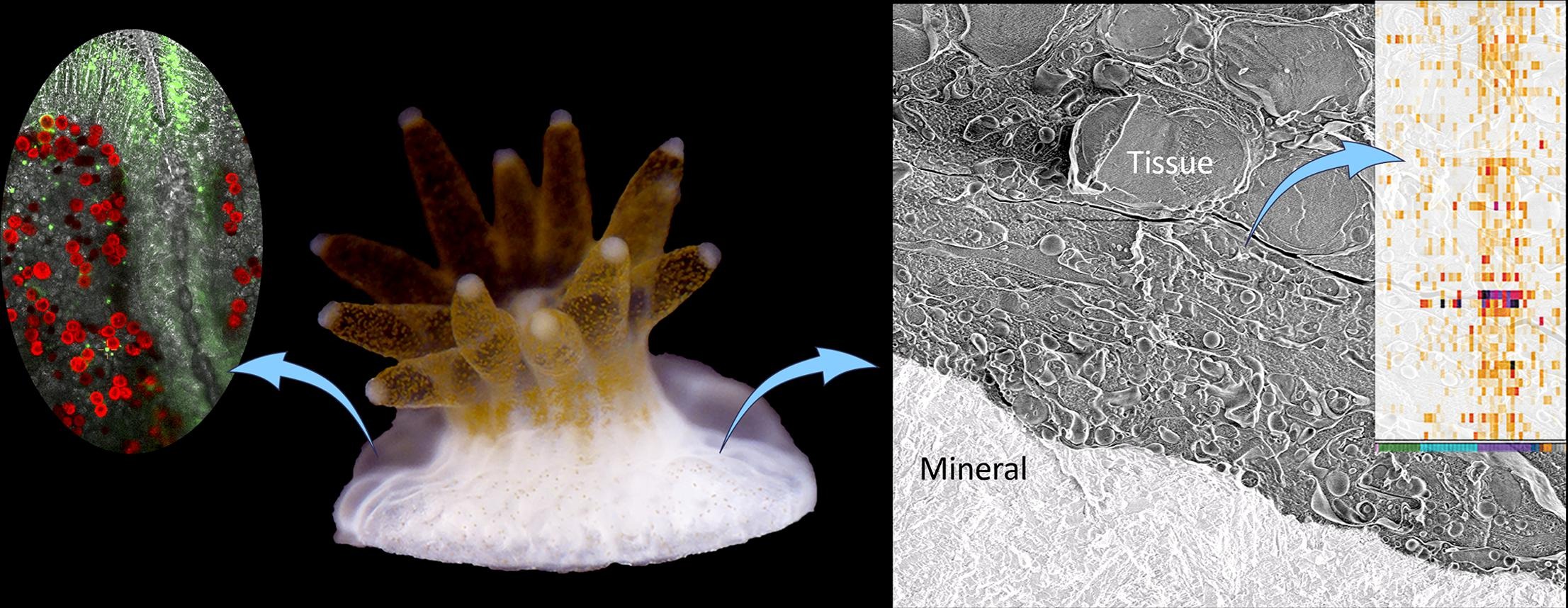
Stony coral exoskeletons build the foundation for the most biologically diverse marine ecosystems on Earth, coral reefs, which face major threats due to many anthropogenic–related stressors. Therefore, understanding coral biomineralization mechanisms is crucial for coral reef management in the coming decades and for using coral skeletons in geochemical studies. This study combines in–vivo imaging with cryo-electron microscopy and cryo–elemental mapping to gain novel insights into the biological microenvironment and the ion pathways that facilitate biomineralization in primary polyps of the stony coral Stylophora pistillata. We document increased tissue permeability in the primary polyp and a highly dispersed cell packing in the tissue directly responsible for producing the coral skeleton. This tissue arrangement may facilitate the intimate involvement of seawater at the mineralization site, also documented here. We further observe an extensive filopodial network containing carbon-rich vesicles extruding from some of the calicoblastic cells. Single-cell RNA-Sequencing data interrogation supports these morphological observations by showing higher expression of genes involved in filopodia and vesicle structure and function in the calicoblastic cells. These observations provide a new conceptual framework for resolving the ion pathway from the external seawater to the tissue-mineral interface in stony coral biomineralization processes.